Hyperspectral Imaging: Tracing Its Historical Roots and Technological Advances

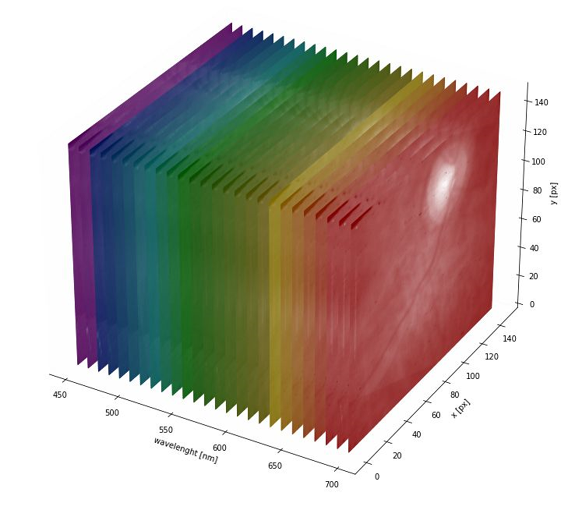
Mantis Photonics’ patented hyperspectral imaging technology has proven applications in retinal imaging and diagnostics. This article focuses on the emergence and history of this breakthrough technology.
Hyperspectral imaging has its roots on remote sensing and earth observation, where it was initially developed to study the Earth’s surface and atmosphere. The technology emerged in the 1980s and 1990s, driven by the need for more detailed and accurate information about the environment.
The first hyperspectral imaging systems were airborne sensors mounted on aircraft or satellites. These systems collected data across a wide range of the electromagnetic spectrum, including visible, near-infrared, and shortwave infrared wavelengths. This allowed for the identification and analysis of various materials and substances based on their unique spectral signatures.
Such information is valuable in many fields. For example, in agriculture it is used for assessing the health and yield of crops, monitoring the soil moisture and nutrient content to optimise crop management practices and improve crop yields. It is also used to monitor changes in land use, vegetation health, and water quality over time, which allows detecting early signs of ecological degradation and tracking the effectiveness of conservation efforts.
One of the earliest and most notable hyperspectral imaging systems was the Airborne Visible/Infrared Imaging Spectrometer (AVIRIS), developed by NASA’s Jet Propulsion Laboratory in the late 1980s. AVIRIS was designed to collect data in 224 contiguous spectral bands, providing unprecedented spectral resolution for remote sensing applications. Several other earth observation programs, like, HyperScout and PROBA-V mission carry hyperspectral imaging cameras.
As the technology matured and became more accessible, hyperspectral imaging found applications in various fields beyond remote sensing. In the medical field, researchers recognized the potential of hyperspectral imaging for non-invasive disease detection and diagnosis. Dermatology is the most obvious example. Hyperspectral imaging is used for non-invasive diagnostics and detection of skin lesions at cellular level. A major application is differentiation of skin lesions from melanoma without the need of a biopsy. Unlike visual examinations which can be subjective, HSI provides quantifiable spectral data that can be used with automated systems for precise diagnosis.
One promising application is the use of hyperspectral retinal imaging for detecting neurological diseases, such as Alzheimer’s disease. Mantis Photonics AB, specialising in hyperspectral imaging solutions, has developed a retinal camera capable of capturing high-resolution hyperspectral images of the eye’s fundus. Changes in the retinal tissue can reflect pathological processes occurring in the brain.
Hyperspectral retinal imaging can detect subtle spectral changes in the retina that may be associated with the presence of amyloid-beta (Aβ) deposits, a hallmark of Alzheimer’s disease.
Mantis Photonics AB’s retinal camera uses a snapshot hyperspectral imaging technique, which captures the entire spectral and spatial information in a single exposure. This allows for rapid and non-invasive imaging of the retina, making it suitable for clinical applications.
By analysing the spectral signatures of the retinal tissue, researchers can identify patterns or biomarkers associated with diseases like Alzheimer’s, Age Related Macular Degeneration and Choroidal Naevus. This could lead to earlier and more accurate diagnosis, as well as monitoring disease progression and response to treatment.
With continued research and validation, we at Mantis are developing the combination of hyperspectral imaging and retinal imaging which holds great promise for advancing our understanding and management of neurological disorders.
