Choroidal Naevi Breakthrough: Navigating Success with Hyperspectral Imaging Studies

Choroidal Naevi are pigmented lesions observed beneath the retina, in the choroid region of the eye, similar to the pigment spots you can see on your skin. While less common than their cutaneous counterparts, skin naevi or moles, they are observed in about 10% of the general population.
Choroidal Naevus, generally, doesn’t cause any symptoms. However, if left unchecked, it can potentially progress to choroidal melanoma. A serious form of malignant cancer of the eye that can spread to other parts of the body and leads to high mortality. This potential link emphasizes the role of early detection and proactive monitoring for timely and appropriate management of the condition.
In our latest article (here), we demonstrated the application of Hyperspectral Imaging in detecting naevi. Contrary to previously used HSI in infrared spectrum, this is the first study to demonstrate the applicability of HSI in the visible spectrum.
Superior detection with higher wavelengths
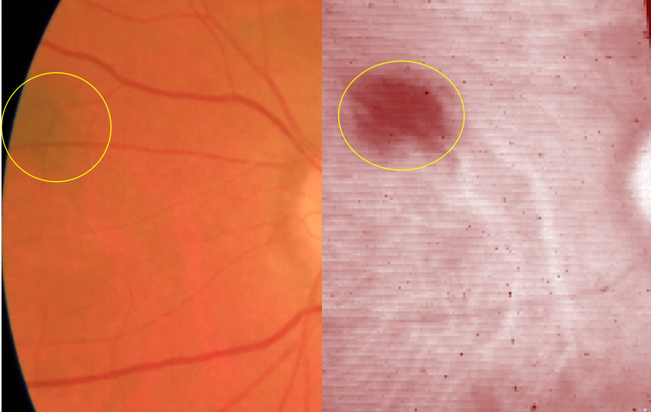
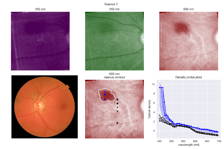
We demonstrated a rise in naevi detection and characterisation between 550nm (the colour humans see the best) and 690 nm at the edge of the visible spectrum.
The following images show how the contrast improves at higher wavelengths thus improving detection.

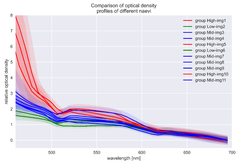
Hyperspectral imaging can distinguish naevi in different clusters
Based on our measured spectra, the naevus could be categorised into 3 clusters (high, middle and low). We hypothesize these clusters correspond to different amounts of vascular density and a component called lipofuschin. As both are risk factors for the naevus to become malignant this needs to be considered when diagnosing melanomas.

Choroidal Naevi are generally diagnosed during routine retinal check-ups or while diagnosis for other eye conditions. However, they pose the serious risk of developing into malignant melanomas with high mortality rates. Hence, it necessitates proactive follow ups. This first ever study for detection and characterisation of naevi with hyperspectral imaging in the visible optical spectrum, simplifies the detection and follow up process. This, once again, validates Mantis Photonics’ Hyperspectral Imaging technology as a valuable tool in the early detection and patient care.